 UPDATE! October 1st, 2023: This version of the website will no longer receive updates. Please transition to the new website for the best experience.
- Lesson Details
- References
- Assignments
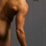
In this series, artistic anatomist Rey Bustos brings you a fun, unique introduction to the anatomy of the human body. In this sixth lesson of the series, Rey shows you the anatomy of the upper and lower arm. Rey will begin by lecturing on the blackboard, breaking down each bone, muscle, and tendon of the region.
Recommended Course Material: Rey’s Anatomy: Figurative Art Lessons From the Classroom
30299 views
Reference 3D (6)


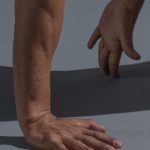
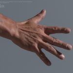
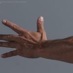
Reference Images (23)


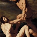
Daniele Crespi, Cain Killing Abel, 1618-1620.
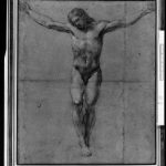
Domenichino Bologna, Christ on the Cross, 1581.


This lesson has Assignments. Subscribe now
Free to try
-
1. Lesson Overview
50sNow playing...
Watch the whole lesson with a subscription
-
2. Learning Recommendation
24s -
3. Bones of the Shoulder and Upper Arm
15m 15s -
4. Muscles of the Upper Arm: The Brachialis
14m 58s -
5. Muscles of the Upper Arm: Biceps brachii, the Triceps
15m 55s -
6. Muscles of the Scapula
13m 41s -
7. Raised Arm View of Upper Arm Muscles
13m 52s -
8. Diagramming Key Areas over Photo References
8m 27s -
9. Ecorche Muscle Drawover on Male (Model: Rajiv)
17m 57s -
10. Ecorche Muscle Drawover on Female (Model: Catherine)
16m 11s -
11. The Forearm: Flexors and Extensors
16m 56s -
12. The Forearm: The "Twins": Brachioradialis, Extensor Carpi Radialis Longus
17m 13s -
13. Diagramming Key Areas/Muscle Drawover Over Photo References (Model: Rajiv)
17m 5s -
14. Diagramming Key Areas/Muscle Drawover Over Photo References, cont.
6m 31s -
15. Assignment
24s

