- Lesson Details
- Transcript
- References
- Instructor
- Iliya Mirochnik
- Subjects
- Drawing
- Topics
- Anatomy
- Mediums
- Pencil
- Duration
- 2h 56m 35s
- Series
- Russian Drawing Course 3: Anatomy
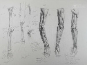
Now it is time to study the anatomy of the leg and arm. In the study of the leg and arm, we are going to focus on viewing the form from as many angles as possible, approaching the study almost like a sculpture. Because these are cylindrical, almost tubular structures with muscles that spiral, we simply won’t be able to understand how some of these muscles look without constantly turning the arm and the leg.
In this lesson, we will identify and draw the skeleton and muscles of the arm.
Students are encouraged to work from the NMA reference images and 3D viewer included on this page*.
Join Ukrainian-born artist Iliya Mirochnik as he passes on a 250-year-old academic method preserved at the Repin Academy in Saint Petersburg, Russia and seldom taught outside of the Academy and never before on camera.
The Russian Academic drawing and painting approaches were uninterrupted by the modern art movements that transformed representational art in the West, and as a result, they provide a unique and clear lineage to the greater art traditions of the past. As a powerful approach that is both constructive and depictive, it combines the two methods that prevail in contemporary representational art.
In these three drawing Courses, we have set out to condense the entire program, spanning over eight years into a logical, step-by-step procedure. We have made improvements and added resources and exercises to explicitly drive home the concepts that are required to work in this approach.
We have also structured the course so that it is not only useful for professional and experienced artists but also artists with no drawing experience whatsoever.
In the last part of our Russian Academic Drawing Course, Iliya brought together the knowledge we learned about the head and neck in order to complete a fully rendered portrait. In this next part, Figurative Anatomy, you will undertake a new challenge: the figure.
In order to draw the complexity of figure we need to study all the anatomy that makes up the surface form of the pelvis, ribcage, leg and arm.
The New Masters Academy Coaching Program directly supports this Course. If you enroll in the coaching program, you can request an artist trained in the Russian Academic Method including Iliya Mirochnik himself. Click here to enroll in the Coaching Program.
Materials
- Graphite pencils
- Kneaded and Hard Erasers
- Sanding Block
- Utility Knife
- Roll of Paper, Smooth Sketchbook paper
- Easel
- Light source
* Reference material is only available for premium subscriptions. If you don’t have premium access to the reference, you can pause the video when the reference is shown.
Discuss this video in the forums!
Discussto the study of the bones and the musculature of the arm.
seen a little bit
of the -
of the head of the humerus, so why don't we start there.
And make sure we have it right here. So
in order to kind of understand
our proportions here,
why don't we kinda try to analyze in the same way we did with the leg.
So with
the leg we were able to find the halfway point. Here the halfway
point's a little trickier. So
just gonna get the bone in there.
The entirety
of the humerus, followed by the two bones
that you see down there, the ulna
and the radius. So I'm just gonna
broadly place
them. And underneath, where you see
the hand,
I'm going to place this area
of the carpals.
And so the proportion that
I'm going to tell you involves -
it's not as clean cut as the one in
the leg. So right here we have
the actual sort of main area of the articulation,
movement of the
bones of the
lower arm, the forearm
where they come in contact with this joint
on the humerus. And so
in general it's -
we can say that from
the end of the carpals to the
top of this area
is equivalent.
Let's see if I have a pencil long enough.
(drawing)
Actually I have my
needle.
The proportion
is approximately equal so
if it's longer I tend
to take either a long pencil or two shorter ones,
establish that proportion. So from the end
of the carpals to the top of that area
and then from the bottom of that part to the
top of the head of the
humerus is pretty much equal.
So that should keep your proportions, for the most part, in check.
Now of course you can't
really pick this area out
but there are certain points on the arm that you'll be able to
explore that will give you
the points that you need. And we'll talk about them in a moment.
So we can
kind of establish pretty much a halfway
point from the bottom of the
carpals, take that proportion
to the
little protrusion that you see right here, which is very
important because you really see it on a human arm.
Right in there. So
we could say, for the most part, that that
is approximate halfway
point. So I'm actually going to pull this area up
just a bit.
(drawing)
Okay.
So before we start to figure out what things are named
let's get
familiar with the shape of the
bones themselves.
(drawing)
So here we have our
humerus. I've kind of enlarged a little bit the
areas of the joints so that they're a little more obvious.
(drawing)
And let's
go and sort of understand what's
happening here and here you have two parts.
You have one part
that has a groove
and has a slightly
more kind of hourglass
structure to it. And then next to it you have something that looks like
a ball. And
these things are quite important. So
before I move on
let's give names
to some areas. So up top here
we have the head of the humerus.
(drawing)
And the part that fits
into the glenoid fossa of
of the scapula.
So this on top is the humerus.
This part on top is the
head of the humerus. And I'm going to
mention these things here,
these protrusions,
which you can see
and that kind of
an empty space in between them
because that is going to be important in the future.
Now, I don't think we're gonna really cover it
but I'll explain what's happening there. And this part right here is called the
greater
tubercle. And this one, as we can understand, is called the
less tubercle.
(drawing)
And that part in between them is the one
that we're gonna need to focus on.
So, I think we can already get into
figuring out some of the tonal aspects of it.
I think we don't need to work exclusively
in outlines.
So you can see the actual the sort of the body
of the bone kind of swerving in
at this point. We're gonna have a better view of it
when we turn it.
But then you see this area sort of opening up a bit.
And that's sort of what's beginning to happen
right there. And you can see that our terminator
on here is quite helpful in showing that, along with a little bit of a
core shadow and a cast shadow onto this part that's opening up.
(drawing)
If we're already putting in the shadow we might as well do that here.
(drawing)
Some of these half tones
to get us a little closer to the truth. The top plane
right there and that actual sort of the head
fits into the
glenoid.
There we go. Okay.
So here's what we have down here. We have this
opening up of this area and it begins
to
not just open up but you can also
see sort of a change in plane, sort of in the opposite
of what we had before. And there's a little bit of a
cavity inside there, we don't need to worry about it.
Though of course
it has a name and all that. But I'm only gonna try
to keep this to the most important elements and all of
these anatomical particulars
you can find in a lot of places.
So I'm just giving you a
head start on them.
So we have already spoken a little bit
because we sort of established this as the halfway point here.
(drawing)
And what these two things are called, I think
you will be familiar
with what they're called. This one right here
on the inside is called the
medial
epicondyle of the humerus.
And this one right here, of course, is called the lateral
epicondyle
of the humerus.
So that's that. We had a fair amount of
experience with all of those condyles and epicondyles in
the bones of the leg. So let's move
on to this point of articulation.
And this point of articulation, those two parts that I spoke
to you about, that kind of hourglass
and the ball right here.
And
their names are, the ball is called the
capitulum.
And the hourglass
is known as the trochlea.
(drawing)
And we can add a couple
of half tones on it.
Okay.
So here's what's happening here and this I think
is quite exciting.
Sort of attached - or sort of connected to -
is the head of or
that's not - connected to the trochlea is
a bone that goes by the name of
ulna.
I'm gonna overlap it right here so you
can see the articulation.
And I'm going to
take it down to
its head, the head is at the bottom
in this case
(drawing)
on the pinky side of the
the hand.
And if you can just take a look
at
this particular joint, then you can already see
what the movement is
between the trochlea and the
ulna. And you can see that it slides up into here
And this part that comes out
goes into that opening over there.
So it moves
the arm up and down.
So this clearly moves us
into this other area. So this is the ulna
and the
bone that articulates off of the capitulum
goes by the name of radius.
(drawing)
And the head of the radius is up here.
And there's this
little part sticking
out known as the radial tuberosity. Don't need to
worry about it.
(drawing)
And the radius
is -
I'll take it all the way
down to the carpal
bones of the hand.
(drawing)
And you can see that there is a gap
between them. Now
of course there isn't a gap, there is a
membrane that fills in that gap
and is the area of
certain muscular origins. But we're not
going to worry about that.
Just trying to understand what's happening here.
And here as well, I'm going to just continue
(drawing)
with some of the tone.
And right here you can
see a little bit of a protrusion underneath here.
And it's
known as the styloid
process
of the radius. But there's also a tiny
one right here which we can't really pick up on the
ulna, which is
is also known as the styloid process except of the ulna.
(drawing)
And so here we have it so I'm going to
add a little bit of tone in some of these areas
(drawing)
because if we spend a little bit of time on the bones, it definitely prepares
us for what's to come with all the muscles
on top.
(drawing)
And then some of these half tones coming
off of these areas of shadow
we're just gonna help really give us the
(drawing)
the forms of these bones.
Alright.
And then down here
(drawing)
we have the
the carpals. Which consist of essentially
two rows of bones,
making up eight bones in total
but we will not be discussing them in detail
we will be talking about them
in the advanced course that is to come in which we cover
the hands and feet in depth.
(drawing)
But so...
So we're gonna call those
the carpals. And if you
remember the equivalent of the carpals on the feet
are called the tarsals.
But these things are quite close.
And then just like
with the tarsals, the bones that come right
off of them
right here, making up the palm and the
back of the hand, in this case the palm,
are the,
you guessed it,
the metacarpals.
And then you have the
(drawing)
what we call fingers
but what anatomists call
phalanges.
And here, for the purposes of
giving us an idea of
the concavity there, just gonna put it in a tone.
(drawing)
Okay. So there we have
the parts.
So the thing that I do want to add is that since we have
figured out the particular articulation of the ulna
against the trochlea, that movement up and down,
what then is the purpose of the
radius? And so
if you look at what's happening here
you have a ball and this kind of -
(drawing)
kind of like a
small cup underneath it
and the movement here
is like this. It moves it around.
(drawing)
And so there's a particular terminology here that explains this. And that
terminology is
for what you see here
with the palms up, the name is supination.
(drawing)
Which applies to
to what we're seeing here.
So now let's observe what
happens when we rotate the radius
all the way. And see so now the back of the hand
is in front of us.
And so why don't we, right here,
we do a quick -
that is supination. Why don't we do a quick sketch
of that. We don't need the
bone on top, we don't need the humerus, but
but we're going to get our
medial epicondyle, our lateral epicondyle, our capitulum,
our trochlea right here.
(drawing)
And then
let's
start with the ulna again and take it all the way.
(drawing)
And see
as the ulna is rather straight.
(drawing)
But now
let's add the radius
and we can see how the radius has a certain
curvature.
And even an angle.
(drawing)
So
I think we're gonna need a little bit of our tone here to
illustrate this point a little clearer.
(drawing)
Gotta clean up that
line and make sure we get a clarity there
on the head of the
radius. We don't need to be overly descriptive
we just need to get the point across.
(drawing)
And here we have our - you can see clearly the styloid process.
This is also a great opportunity to now quickly review.
Like there's a lot to be said for having
a review right after
right after you
learn a thing. So we have
the styloid process here
and the head of the
ulna.
(drawing)
And there it is. And then we have some -
a shadow being cast from the trochlea
onto
the
ulna itself.
(drawing)
Okay and then
the carpals
in a few rows. The metacarpals
and then the
phalanges underneath. Okay. Now what this
is called pronation.
(drawing)
And so there is a point
in between. And that
is going to be the arm that we will be
working from in the écorché.
So I'm going to turn it slightly so it
becomes clear. And that is
when the
palm is
turned towards
the body. And what that is called -
this is called is
demipronation.
Or half pronation.
And of course they're are all these
degrees in between. It's not only
up, half, and around.
But this is going to be the position in demi-
pronation that we're going to be working from in our écorché.
And so the thing to think about
in this case is the particular angles
of the angle between
the upper arm
and then lower arm. So if this is the
humerus and this is our ulna,
that we know is relatively straight, though it kind of
there is a slight angle outwards there.
A supination. But to make it clear, this diagram
you can see
that the particular movement of the
radius is actually gonna move this,
really affects the angle here.
There's a movement outwards.
And then once we attach
all of the muscles, it becomes even
more clear. And so
as soon as we alter that and we have the humerus
and we have the ulna
and then we have the radius.
You can see that in as
the radius turns from a
position of supination into a position of pronation
that angle
(drawing)
that angle moves closer to a straight line.
Or in this case, you know,
a hundred and eighty degrees.
Okay. So let's keep that in mind. And
the final thing that I would like to talk about is of course an important element that we can
only see from the back, we can see it right here.
And that is the elbow.
And so if we simply draw the elbow
and you can see right here the
medial epicondyle.
And you see the opening here too in between
here we have the
other side, we have the -
the
lateral epicondyle.
And right here you see the bone of the elbow
(drawing)
which is this large part that sticks out.
If you take a look and take it all the way
around
you can see that this is the ulna
because right here you can see the
trochlea.
And what this is called
this
elbow protrusion
(drawing)
is the olecranon. Olecranon.
And
it - you can see
it's like, it hooks on
to the trochlea here.
And this part fits perfectly
into
this area right here
which goes by the name of
the olecranon fossa.
And we've already encountered that. It simply, it means
a hole of a sort.
A depression.
(drawing)
And there
is that. So let's
try to just make sure we get an idea of
what the structure of the olecranon is.
It's kind of - you can think
of it a little bit like a rectangular prism, a little bit like a cube.
And parts up here. It's got
pretty clean and
very sort of
precise changes in
plane. It's got a
top plane, a front plane, a side plane, all of the stuff that we see here.
With of course, we can also see a little bit of that kind of opening.
up there. Okay so
I think that, for the most part, concludes all that we need
to understand
before
we begin our analysis of the muscular
anatomy.
arm. And now that we have an idea of what's
going on inside, let's approach
this in a similar way that we did with the leg,
working
from multiple viewpoints. Here however
we're not gonna do the upper arm and the lower arm separately.
I think we're able to take it in one go.
So
here we have our upper arm and you've noticed that
the cast here is cut at the deltoid so
we're not actually, we don't actually get to see the head of the
humerus.
And I'm going to
slightly increase the
the volumes of just some of these muscles.
Just a tiny
bit so we can see them
(drawing)
but right now we're just getting acquainted
with what's happening here, we're focusing
much more on just what
this looks like.
(drawing)
And that would be a point I mark because that's our olecranon.
And then
right here we have
our lateral epicondyle
and underneath it
a tiny point - you can't really see the whole thing but that is
the head of the
radius. So let's continue.
So if
we know that this right here is the olecranon, we can
continue with it
and arrive at the head of the ulna.
And then we can take a proportion.
(measuring)
So from the olecranon to
the cut where we have right now -
oh interesting. Seems
about -
seems like from here to here is exactly
the same as from here to here.
Not bad.
I like what I'm seeing.
(drawing)
And so
here we have a group of muscles up there,
we won't get to naming them just yet but we're going to follow along.
We know that this demipronation.
We also know that the radius
is going to be right here
in profile
view essentially. So we could place it almost in
its entirety, that would be the ulna.
(drawing)
And there we have it.
So we can move into our
metacarpals in there. And
the - you can see the metacarpals, the carpals in there but
then you can see that slightly bulging area of the
heads of the metacarpals.
We complete the back of
the hand.
(drawing)
And there we have it.
So why don't we talk about a few
things that we can pick up here.
So -
and let's do that before we move on.
Here we have the deltoid closer
to its insertion. We've
seen the top of it but we have not seen where it
inserts at about halfway into the humerus.
And in the -
in the back here we have the triceps.
And the triceps,
let's start putting some names down,
we're aware of
the deltoid. The triceps
so called
because they're three pieces and we'll be able to see them
a bit more when we turn the -
but here the part that you see,
and you have seen a part, you've seen the long head, which
we can see right here I
think. Yes. The long
head of the triceps, the one that -
the one that you saw come out in between
the teres major and teres minor.
(drawing)
And
here we have the lateral head.
So the long head and the lateral head are the ones that you can see.
(drawing)
And let me see from the other angles
(checking)
Yeah. I think we are going
to get an opportunity to see the medial head on the other side
where it sticks out just a tiny amount
but it's primarily underneath.
So if you can imagine
here we have our -
the back of the
humerus.
And here we have our medial epicondyle.
Here we have the
the medial head,
we have the lateral head and on top here
is the
long head, the one that
comes in
between the teres major and minor.
And there's a tendon that connects them
in between
that connects essentially all of them
really. And then
moves into and connects to the olecranon.
So here's what we have right there.
(drawing)
Now here is
a muscle that's
attaching right to the humerus
in the front, know as the
brachialis.
And brachi means arm so
that word might keep appearing.
(drawing)
So here
on top of that is the
muscle that I'm sure most people are already aware of and it's known as the
biceps.
(drawing)
And we can't really see what's happening from this angle
so we're not going to
really talk about it right now.
I think as we turn the arm there'll be a lot
more that we'll get to explore.
the lateral epicondyle.
And there's an entire group of muscles
that originates at the lateral epicondyle
and attaches to the back of the
hand and the fingers as well.
And that group is known as the
extensor group.
And
here you can
clearly pick them out as almost
sort of a solid area.
Now I'm not always, not absolutely,
but for our purposes, let's try to group them
even though I will tell you what they are.
(drawing)
There's also a group that originates at the
medial epicondyle and attaches to the
palm side of the hand. And that group is called
the flexor group.
And now they're aren't any that we can see from this
angle but as we turn the arm, you'll begin to
see where they are. So
(drawing)
right here I'll just - I'll quickly
point out there is this sort of triangular
muscle, sort of coming out
of the olecranon, coming off of the elbow, known as the anconeus.
(drawing)
Anconeus.
So here's a group of extensors.
So we have
for the most part covered
all of the things that we are able to pick up here, not really getting into the hand
though it's hard to kind of avoid the hand
entirely
despite my efforts to kind of
make this as clear as possible, mainly because most of the attachments
of all of the muscles up here are on the hand.
But we're more concerned with the sort of the structural
qualities here so I'm
more interested in just pointing out where things are
and how they affect the structure of the arm.
Okay so and then
up here you have two muscles that are sort of
in between the
extensor groups and flexor group on the other side
and
they
originate from
this edge right here
above the lateral epicondyle. And they are called
(drawing)
brachioradialis
(drawing)
and extensor
carpi
radialis longus.
(drawing)
And you can see them right here. And they too can for the most part
be thought of as a group. And their tendons
continue
to the ulna there. Oh sorry to the radius
right here. And their
tendons extend to the radius
over there. That too was a note to the editors. That's the correct one.
Okay. So
since we've started to
say what these things are called, why don't we continue.
So now that we're here we've
arrived at the group that we've already briefly talked about,
the
extensor group and there's
a part of the extensor group that you
up here can think of as part of
the extensors but down here
it sort of moves off as you can see and attached itself more
to that kind of group in between the extensors and the
extensors and the flexors. And that
muscle is known as the extensor
carpi
radialis brevis.
So it's like the one that we
covered
but this one right here is the short one.
So now we really get to our
extensors right here. And here
we can get a little more specific but I think
this'll be, I think enough.
Is
a muscle that's a lot
like the one that we saw in the leg
continues and
extends to all of the fingers
excluding the thumb.
(drawing)
It is called extensor
digitorum.
Kind of like the other one was called too
except that was a long one.
It had a long attached to it.
So right here
is another one of the extensors
(drawing)
and it is called
the extensor
(drawing)
carpi
ulnaris.
And now we do get
(drawing)
something happening right here. You do get a little bit of a
mass in there and it's
kind of helpful
but it's a muscle that's -
it's part of - it's a bit more
inside
(drawing)
but it kind of - you can kind of -
I think it's helpful because in between the extensor
carpi ulnaris and the
already sort of part of the flexor group
and we'll look at it, the flexor
digitorum
profundus.
And it's deep inside so that's what
that word means. It's profound.
(drawing)
And so
you can see the olecranon
coming out in between these two but you also see the
head of the ulna coming out in between them as well so they kind of
they help in guiding you to see where that ulna
is inside.
a few more here
(drawing)
and they pertain
to the thumb. Because as you
are already aware, the extensor digitorum
does not -
does not move the thumb
at all. So because we have
flexible thumbs, there's a lot that goes into making that happen.
So here's two muscles
that are really going to help
with that action.
And they are called the
abductor
pollicis longus
and pollicis is the
thumb. And the one underneath is
extensor, this one right here,
and the one underneath is the extensor
pollicis brevis.
And so
they create -
they create a certain amount of relief in that area so that's why
it is important to cover them.
And they have two tendons that play an
important role and there's -
there is the tendon here that we should cover.
And as soon as
we - as soon as we turn the arm
we're really going to see these tendons
play the
role that they're going to play so that tendon
belongs to a muscle that's in there, known as the
extensor
pollicis longus. You guessed it.
so there's a long one and a short one and the short one happens to be on the
outside more. So that about covers it
for what's happening in this area
so I'm going to turn the arm
so that we get a view that's somewhere in between the anterior
and the
medial.
(adjusting)
And
let's begin.
So since we already have all of this
we might as well use it.
(drawing)
Hmmm, I think we're getting a -
okay yeah, that's the
brachialis in there.
(drawing)
And here
you can see it so clearly is the
medial epicondyle of the humerus.
(drawing)
So
we're just going to lay it in
and here you can already see those tendons
that we were talking about. Those
tendons
going to the thumb
from the
abductor pollicis longus.
The extensor pollicis brevis and
this tendon right here, coming
off of there is the extensor pollicis
longus. So - and they're really creating -
they're sort of playing a major role in that gesture of this
demipronation. They're kind of exaggerating it. It's nice.
(drawing)
Okay so why don't we begin
with stuff that we already are aware of. So right here
we already know is the
sort of bottom portion of the deltoid at its insertion.
Its insertion is further inside of course but
that's what we have here.
Now
(drawing)
hmmm that's interesting that they have this here,
that's awesome.
There's actually a little bit of cut which
I'm going to place here because we do know where this
is - that is the like
the part that's attaching on the pectoralis
major. So we can imagine it coming over
this and coming out
of this
area. So that's pretty cool.
On the other hand it's quite nice that it's not there so we can see
what's happening with the biceps.
(drawing)
And so here
we have the biceps. So called because of the
two heads, remember the
triceps had three heads and the biceps have
two heads. And here is the brachialis, which we had
right here. And we see it sweeping
in there and the biceps is on top of it.
And I'm also kinda just slightly enlarging
everything so that's a little bit clearer.
And so the reason that -
the reason that I mentioned to you this
area right here, that the groove in between
the greater tubercle and the lesser tubercle
is because if you imagine again
where we have our
(drawing)
our
scapula
(drawing)
then one head
of the biceps begins at the
coracoid process.
So yes, the short head of the
biceps begins from the coracoid process, that part that sticks out
right above the glenoid fossa,
while the long head
is sort of a tendon that comes from right above the glenoid
fossa and wraps around the head of the humerus,
passes into the groove, and then
here
we have the deltoid where both of those parts
combine. Underneath it all
is the brachialis right there. So
that's what we're seeing here. Now what we don't see the
head of the humerus, which is a shame. But we do see
the bicep splitting up a little bit and you can see
the short head right here and you
can see the long head right here and then they kind of become
a single thing. And then
they connect into
this part that sticks out right there, the
tuberosity of the radius as well as
with this sort of
tendonous area, the aponeurosis of the
biceps right on top into the
fascia of the lower arm.
And we see it all here which is
wonderful. Now what -
now what we need to make sure we see
here actually we have a little bit of brachialis as well we can kind of just
imagine it in its entirety. We have the humerus right there.
And here is that
part that I spoke about, the
medial head that you would be able to
see right here, right underneath there,
coming out from under that
tendonous area. But there is the
medial head of the triceps and right here on top of it is the
long head.
(drawing)
There we have it. Here we have a small muscle
(drawing)
known as the coracobrachialis
because it begins at the coracoid
process and attaches to the humerus, the
brachhia, the arm.
And here we have a few things that are
the - it's hard to
place them mainly because we don't have
the origin really. Because we don't -
because we don't have like the top,
top of the humerus here.
Because these would continue higher but here you do have
your teres major seen here,
which you would not see of course because of the covering of the
pectoralis as well as the infraspinatus and
all that. And you would
be able to see the infraspintus here because it connects to the back of the
head of the humerus and all that. So we're not gonna be too concerned with that but just keep in mind
that the head of the humerus is an important point.
Okay. So let's move on
and see what else we have already spoken about. So here we have
the group that we have
discussed already.
And you can see it just a little,
you can see the brachioradialis
right here and it's fairly large and has
and is very prominent and it's always starting
a little bit higher than your lateral
epicondyle. And then coming out from it
is the extensor carpi radialis longus.
(drawing)
So that's what we have
in this area.
flexor group coming off
of the medial epicondyle.
So there's a lot happening there
but
let's talk about the ones we really need.
So right here
coming down into there
a muscle of considerable size, enough for us to
pay attention, right there, and it's -
we can't see its tendinous attachment into
the carpals but
this right here
is flexor
carpi radialis.
(drawing)
Now
I'm going to write them in a strange order.
So bear with me.
And so the group
here - you can group them for
the most part -
so the group that comes afterwards -
and I'm going to group them - is
a combination
of two muscles right here
and it's hard to see them - they overlap
from this angle in some places and they continue
all the way into the palm
and the group consists
of this muscle up close which is a
a long one. And just sort of attaches
into the palm, is known as the
palmaris longus.
And then right behind it, we can't see it, I mean if you walk around
you'll see it but
just think of them as a group, is the
flexor digitorum
superficialis.
So we've already seen the
flexor digitorum profundus right here.
That's the one inside and the
superficialis is the one on the outside.
And so then we have
another one that's quite large
and also quite important and is
already kind of on its own. You can't
totally group it in with the rest of them.
And continues here.
(drawing)
And is called the
flexor carpi
ulnaris.
(drawing)
So we've seen the extensor carpi ulnaris
right here. And this
one right here is essentially its opposite,
its flexor. Which you could combine into a group
with the flexor - you combine the flexor carpi
ulnaris and I'll add
this here with the
flexor
digitorum profundus.
So
I included it here when talking about
the group of extensors but we know
it's not part of it due to its name and so
I'm going to move it over to the group of flexors.
And I'm going to add one more on top here.
One
more right in there
known as the
pronator teres.
And we've seen teres before it simply means that it's round.
Now is it really
round, how round is it?
It doesn't look very round but
that's what they called it
so we're gonna go with it.
So
before we move on, why don't we
continue with these
muscles and the tendons that come off of it
of the thumb.
And we see this
top tendon right here,
we saw it right there, belonging to the
abductor pollicis longus. And
it attaches right to the base of the metacarpal
(drawing)
of the thumb.
And then underneath it we're gonna continue
and sort of going around
that area, opening up
and in between the abductor
pollicis longus
and the tendon of the extensor pollicis
brevis, which continues
onwards,
you can see the head
of the metacarpal there.
And then we can see
right here
the tendon of the
(drawing)
the tendon of the
extensor pollicis longus. And in between
there's a gap.
Right here
as well.
That's really going to help with
what we're seeing there.
Now right here is another tendon and I don't wanna get too caught up
in them even though they're so important - is another
tendon which is the tendon of the extensor
carpi radialis longus
and they kind of overlap there.
And then we can take all of
this out right there.
(drawing)
Okay so I think
that about covers it. Now I think we're
not going to see anything else come along really so we're
just going to see what it looks like from yet another angle. So
we're gonna turn this again.
We're gonna turn this again.
(turning)
Like that,
(drawing)
so that we can have a better view of the tricep
and a combination
of our extensors and flexors. So let's
lay this in.
(drawing)
We known our olecranon is right there
and we can immediately - so I'm gonna speed this up just
for you to see how if you do have an idea of where the anatomy is
you can kind of lay it in with that anatomy in mind right away.
So here we have our triceps and
don't worry we'll go over all of it. Here we have the anconeus
right here, the ulna itself, and the olecranon, very clear here.
The -
the
epicondyle there, the medial
epicondyle, the lateral epicondyle
the brachial radialis and the extensor digitorum
longus.
Then we have the group
here.
Oh no - okay. So here we have the
brachial
radialis and extensor carpi
radialis longus.
Here we have our large area that we can just
think of as a group of the extensor
digitorum right here.
And with the anconeus right here and
then on the inside, the other side of the
ulna, we have the flexor digitorum
profundus with the ulna popping out.
right there. And then here we have the
flexor carpi ulnaris.
(drawing)
So now we can get into it and maybe
clean things up a bit.
(drawing)
Okay so here
we have the lateral head
of the triceps
in front of which we have the brachialis right there.
(drawing)
Now here you can really see the
long head of the
triceps and just the
tiniest amount of the
medial head right there. Now
we need to cover this with our deltoid.
(drawing)
Oh and here we even have a little
cut away, we can see the posterior portion
and our teres
major up there, we don't need to worry too much about it.
And remember that I'm slightly enlarging some of these
things so that they're a little more human
and not as
cadavary. Though of course
the clarity here is
superb. So here our olecranon
and the common
kind of tendon
of the
triceps
and our olecranon and ulna are right here.
Now we're gonna continue with the anconeus, that sort of triangular
muscle on the like
right on the olecranon and make sure
to be able to really
establish where our lateral epicondyle is. And then
from
above that area, we can begin with the
brachial
radialis, which is beginning to sort of
sweep away from us, and the extensor
carpi
radialis, which you can hardly
see though.
So - but in reality you, from this
angle, you hardly see the brachial
radialis either so this is the importance
of keeping these things in groups.
(drawing)
So here we have the group there.
that tiny
thing we see up there is actually just that top
portion for it sweeps around and other of the brachial
radialis. And underneath it you see the
extensor carpi radialis. That
group that begins from above the
lateral epicondyle. And then we can continue
downwards and we see
the extensor
digitorum
(drawing)
followed by the extensor carpi ulnaris.
(drawing)
And you can see sort of
wrap around there, wrap around the head of the ulna, opening up and you can
see the ulna right there. And remember, we need any
opportunity - we take any opportunity where we can see
the bone. Now on this side,
that opening, is the flexor digitorum profundus
remember that inside part that.
We need - but you can still kind of find as a group
and combine with the group here
of that flexor at the very end
the flexor carpi
ulnaris
(drawing)
into its attachment
at the hand.
And you can just see there's -
so I'm sure - I haven't spoken about it
but you can see that there's this area that belongs
to pretty
much where the carpal bones are. And this is definitely something to talk about
in connection with the hands
but I want to still make sure that
you kind of always try and see where the end
of the radius is, where the end of the
ulna in this area and then
imagine where the carpal bones are and
think of the wrist itself
as having its own orientation, its own angle
apart from that of the arm
and the hand in between.
And so here is what we
have come up with. And for the most
part we've covered a lot
of important material. But at the same time, don't forget
to think of these areas as a group. So overall
even though of course you can
sort of keep dividing them based on what you're trying to
accomplish but think of these things as
the groups of
the extensors, the group of the flexors, that group
in between the two
and the group that
is right here that controls the thumb.
Okay so now that we've outlined
what's happening here
let's try to make this
into a more
structured representation of the
arm. So I'm gonna go here and turn it back
to its initial position where we had it.
There it is.
(drawing)
And let's get
started. So of course we need
to figure out where our shadows are. So we can see the shadow from
the deltoid
a core shadow.
And the cast shadow it's casting on the
lateral head of the
triceps. But then we can continue
and establish the
actual core shadow of the triceps itself.
(drawing)
And take it all the way
down including its tendon, into the olecranon.
(drawing)
Now we have a little bit of
a shadow and this one's not gonna be too
easy to see on people but you can see
some hints of the brachialis
but it won't ever be as obvious as this
right
in there
(drawing)
and then the biceps
are in the front there.
(drawing)
Okay so right here
to our advantage, coming off of the group, that
intermediary group between the flexors and the extensors, or
you can even think of it as sort of
the added on extensors
they're still, to some degree, extensors but
they don't originate from
the exact same place as the other ones and
they stand out a little more so
I like to think of them as a separate element. As you'll see here why
it's nice to think of them as that, mainly because you can see
the shadow, both core and cast
that's in this area coming off of them. So there's
always, from a structural standpoint, you're always
thinking of them as a separate
group there.
with its own changes in plane.
(drawing)
And then
we could make sure to have the core
shadow on the anconeus
which you can also think of as an element that will just emphasize your olecranon.
(drawing)
And then we can think of
the group of the extensors here. So if you imagine this without
this line, cutting them apart
into their individual muscles
(drawing)
you will see them right there. So you think of this
as this roundness. And then here we have the
extensor carpi radialis brevis
that is going to open up there a bit
and here that tone that we
see there on its side plane is what's going to help connect it
with this
group
of the brachial radialis and the extensor carpi radialis longus.
I'm sorry, I know that there's a lot here and
these names are terribly long and
but I think even the way that I'm
placing this on paper right now is helping to kind of see how we can
think of them as a group. And we're probably gonna
lose that a bit as we always tend to when we get into
the details
but nonetheless. So
right here,
is, you can see from a
tonal standpoint you can really see
that sweep inwards that's coming off
and of the radius right there and onto
this area of the carpals and then
that pull out right here of
metacarpal bone of the index.
You can see it there in that opening
in between one of the
tendons of the extensor digitorum.
(drawing)
And the -
and the
extensor pollicis longus.
And you remember that
right here is that small tendon, we had it
right there, let's put it in here, is the tendon, the attachment of the
extensor carpi radialis longus.
And you can see even closer right into
that head of the
metacarpal of the index right there
is the tendon attachment of the extensor carpi
radialis brevis right there.
How important is this? It is when you're working on the hand
and I'm only focusing on these here mainly because
they lead to
a landmark right here, that opening of the
metacarpal of the index.
It plays a role there in a sort of a point you need to take
to keep your eye out for on the hand.
(drawing)
Okay so
let's do a little bit of work with some half tones
before we move on
and now we can make sure to isolate
the - in the group there - to isolate the
extensor digitorum. Now if you look you might see this extra
element there, the extensor digiti mini
and it's a part of the extensor digitorum
that sort of like splits off and
sort of attaches to the pinky. We
don't really need to think about it.
But right here we still need to think of their overall mass
Right. So that's what we're going to
do.
(drawing)
And we have a cast
shadow from the extensor group onto the anconeus.
(drawing)
But then right here
we can use the extensor carpi radialis
brevis to get a little bit more roundness to this
intermediary group because the extensor carpi
brevis is going to be sort of a change in plane. You can see it happening right here. One,
two, and then a third in there.
(drawing)
And then we move up and right at the area
right at the area of the
lateral epicondyle
is when there's a major change in plane happening
in this group.
And this area on top begins to catch more light.
(drawing)
And then up here
like with some
of the things that we've already seen, we have an
opportunity to add a little
extra on the
side plane of the lateral head of the
tricep, sorry. So
we can get this into a half tone
(drawing)
And that's what we have right there
Now,
I like a cast shadow,
I like when it's giving us more form
(drawing)
and also I need to make sure some of these important
terminators are
in place.
And then we can do
a little bit more work here on the deltoid,
(drawing)
making sure we get
(drawing)
the turn,
(drawing)
overlaps,
and our core shadow.
(drawing)
Okay. So I think that's
a good amount of analysis. You can see how now it begins to integrate into a larger
structure, a larger
understanding. And once that happens
it's not as frightening
because all of this is sort of frightening.
(drawing)
So let's move on.
(turning)
Excellent. okay. So here
(drawing)
we also begin to think -
actually so a tiny bit
more right on that, yeah.
Okay.
So let's begin
with this important terminator along the
biceps. And it's honestly
kind of interesting because it's going to
give us the distinction, that sort of cut in between and
you can see this on some people actually that
cut between the long and short heads.
(drawing)
Because we're going to get a little bit of
light on the long head there.
Right here.
(drawing)
And
then you can follow this upwards
to get our brachial radialis which we can see
right here.
And right behind it always
next to it is the extensor carpi radialis longus.
God these names are
so long.
ECRL.
We can abbreviate it but
then we have to abbreviate all of them and then it's - and I think
some people they do abbreviate them.
You can call it ECRL.
That's almost harder to remember I think.
Alright so let's get
in there. Here you actually see a little bit of the brachialis,
the one that you do see inside here.
And we're gonna have to differentiate between some of these things
so that we get a little more clarity on what's happening
there.
But not just yet.
Here let's get that little bit of a change in a plane
on the long head of the triceps. It's nice to see it
and we can get this a little cleaner.
(drawing)
And then
(drawing)
right here on the medial head
we have a little bit of a
shadow with an occlusion shadow or
possibly just a very, very small cast shadow onto
the humerus. Okay.
(drawing)
So now let's get
our ECRL, extensor carpi
radialis longus. Maybe it's not so bad to abbreviate them.
(drawing)
Let's make sure they are -
this is all in shadow. And then here
carefully, right in between our
tendons we also need to place those shadows.
(drawing)
There are muscles here,
the interosseous,
but we're not going to get into them.
But for this the eraser can come into - a small eraser can be really
handy to get some of these things clear.
And to really get those overlaps of those
tendons.
(drawing)
So there they are. It's nice to use a shadow to show them.
(drawing)
Okay.
Now that that's in place, let's just move up a little bit here
so we can get a
tone on the deltoid.
(drawing)
And let's begin to analyze, so here
we have pretty much a very clean change of plane.
This is nice. This is very good. We have a very clean change
of plane on the brachial radialis and kind of in between the brachial radialis
and the extensor carpi radialis
but - and it continues all the way down to here,
we can see this change in plane all the way
into here.
And then from -
from essentially the end of the flexor
carpi radialis,
FCR. I don't know if that's more confusing than not
but is essentially this
change in plane. And of course it's all round but
we're trying to box these things up a little bit
and it does in fact become more of
a box as you get closer to the wrist.
(drawing)
And so
here is kind of a plane on its own. So if we
think of it like this, this is possibly oversimplified
(drawing)
but that's what we have there kind of.
I think we're pretty good and then
we need to make sure there's a proper -
a proper curving there,
a proper curving
in. And actually
I'm going to raise
this a bit.
I'm gonna pull this up a bit more
as well. I think it's more effective
just a little bit. I need to elongate these things I feel
that that's what needs to happen.
So here we are.
Kinda begin to get
a gradient up there because this right here
is that same place, change in plane right there.
And that change in plane, that turning upwards
and also turning around
there is something to capture.
So there's a movement along
our vertical as well as across our horizontal.
(drawing)
Hands are great.
I can't
wait to get more into them.
(drawing)
Okay. So
right here, we need to use that side
plane of the bicep.
(drawing)
And to take a look at Michelangelo, he
pays so much attention to that -
to these tiny side planes of muscles.
They play an incredible role in how he is
able to economically describe a form.
Usually he'd just establish that,
establishes a few of those side plane,s
a couple of overlaps and he's got everything you need.
(drawing)
And then here, from that aponeurosis,
that attachment
of the biceps,
we get a cast shadow
into
onto our pronator
teres.
(drawing)
Okay. Okay.
Just a little bit more right here
to make sure we
get this light
on the brachial radialis
to make sure it's clearly coming out and above
the flexor group.
(drawing)
Alright. Let's take a look at the posterior aspect
and I just turn the
arm here.
Perfect
Yeah.
And let's do a bit more
of the same. And here
we're going to begin with the
(drawing)
the terminator on the long head of the
tricep.
(drawing)
Because that's our main terminator, that's the terminator where the arm
turns finally away from shadow.
So if we were to think of it as a strictly cylindrical
object, that would be our terminator on the cylinder for the most part.
Or close to it. Oh, my apologies.
(drawing)
Okay so - and now
we're going to use the posterior portion
of our deltoid, its core shadows
and all that stuff
to make sure we get those nice, clean cast shadows.
(drawing)
The heads of the triceps.
(drawing)
And then here
on that tendon of the triceps as well as,
making sure that the olecranon really reads
and we're gonna go back and do it of course.
(drawing)
We're really going to go back into it,
as we did with
the leg and the knee, making sure to emphasize
all of our important
landmarks, which if you haven't noticed all happen to be
at the joints on the outer leg.
(drawing)
So here I am continuing,
this is a small sort of minor -
a minor shadow on the
anconeus and then I'm gonna just drop it down here
on the flexor, on the flexor digitorum
profundus
because that's our main one, to follow that terminator
down and about, right to the ulna.
(drawing)
And there we have it. And we can make sure the
rest of it
(drawing)
is clean there
with a little bit of the tendon
of the flexor carpi ulnaris.
(drawing)
Here we actually have the ulna, we must keep that
in mind. Right here we don't have too much
in terms of -
we don't have too much
(drawing)
in terms of that intermediary group, that important group but still
that one in between. But we still
need to show anywhere we can and maybe it could be done more with light
by actually grouping them.
And remember you want to get that group. And see even
the extensor digitorum, up to
a point, sort of continues that movement.
It continues
up here at its origin, it continues
that
rotation. And then of course it splits up.
But we can see it from this angle, which is nice,
and then here the rotation is not the same.
And we follow - so there is some advantage to having
an idea of where each one of these is.
And it's not even that difficult to remember what the names are
because the ones around the ulna, if
they're coming from the inside, from
the medial epicondyles, are from,
in terms of their names, will have
flexor in the name as well as ulnaris.
And if it's - and the same thing applies
for the extensors and then if it's
that same part or a part that sort of like clearly
attaching,
clearly attaching to the radius then it becomes
the radialis. So in terms of what they're called
there is a very clear logic here.
It's just the names are so long, it takes you half and it takes me half
an hour to say them. You might say them faster
almost sure you'll be able to say them faster.
(drawing)
Okay
so the only here
plays a good role here, kind of
gives us some interesting changes in plane. So
from the ulna to the tendon of the
flexor carpi ulnaris is essentially
the side plane of the wrist. And then
going across the ulna,
across the group of extensors is the plane of the
the wrist at the back of the hand.
This is the importance of being very clear
of where things are because of the movement of the
hands and the arm and
because the back of the hand can essentially become the
front of the hand in pronation.
It's still the back of the hand but
that's where one might get confused. So we need to
kinds be as clear as possible
with our orientations there.
And so we do see that happening
here. On the
extensor carpi ulnaris there is a bit more of a change in plane
and so here we're going to need to get
a half tone to show that. And then there's a real kind of beginning
of that wrap around right here on the anconeus.
From the anconeus, from the ulna and the olecranon here
is clearly the movement
to the other side.
(drawing)
And then here,
let's establish some sort of
side and front planes as well.
(drawing)
Because the long
head of the triceps comes
out a little more, it's catching a little more light so we do see
that happening, so the high point of it
in a slightly different place.
So here we're gonna really have to use our light to
get things across the
brachial radialis in there, we're going to need to show that with light as well. And then
in general here the extensor carpi ulnaris
I'm sure we can get
in a general half tone.
(drawing)
And here using that cast shadow,
the light onto the carpals.
Right onto them.
(drawing)
And then here, on the other
side of the
ulna,
we can see the tendon of extensor carpi ulnaris,
right there coming around.
We can even see at a nice, clean point where it inserts. So
we need some of these tendons, don't wanna go too far into them but they are there.
It's nice to know where they are.
(drawing)
And see so I hope I'm not
over - I'm not over
emphasizing this anatomy. I'm trying to keep it to
essentially the absolute
minimum that we need. Now don't let that
scare you at all.
But the whole point is not even so much
being aware of
what everything is called as it is
you can see how having an understanding of where the
placement of all these things are as
really helps
with structuring everything but even
somehow more importantly, figuring out where you need
to place your most important accents. Okay now that we've
moved these along, let's go back and sort of
polish each one off.
and let's see what we can do to complete these
drawings.
Because remember there is
a point in simply
doing a lot of anatomical studies but
think it's not such a bad idea to use them
to also practice the just actual
sort of mechanical technique of drawing as well.
Sort of learning to take them to completion
and
kind of even adding some
interesting stylistic elements.
So here we are. Right
up here, just like we did with the leg.
I'm focusing on the area around
the
epicondyles and
the olecranon.
(drawing)
And I'm going to make sure
these areas
really
are clear and have the right
amount of contrast.
Along with, if you remember, a nice
solid contour in some places.
So
remember the importance of overlap.
And here we can see the anconeus.
overlapping the olecranon.
And then on top here we have the
tendon of the triceps
as well. So these overlaps are really
important. So here too
is that tendon and the
end, as far as we can see it,
lateral head of the triceps.
(drawing)
And right overlapping in there
is the long head of the triceps.
So a lot of information on those overlaps.
Play close attention to them and try to
remember them.
(drawing)
And then to get into the
(drawing)
terminator there as well.
(drawing)
Now here
gonna get some work done on that
tiny change in plane on the brachial radialis.
(drawing)
And right now, completion
of all different kinds
the completion of sort of an academic assignment is not exactly
the same approach that one takes
when completing
something perhaps
a bit more creative. But completion
in general is one of my favorite parts
and I have to tell you that in school at
the Academy, I hated it
and would leave a lot of stuff just incomplete.
Possibly because I just didn't know how so
I kinda skipped that part
even though sort of piece by piece slowly
I would get a little bit closer
to a certain kind of completion,
an academic completion, sort of a clarity of edge and
all of that but I took a while to learn and
I learned it sort of one step at a time so
just keep at it.
(drawing)
So I'm just
going over these
(drawing)
areas in here, we have
the extensor carpi ulnaris
coming right up against the
anconeus right. So we want that over
overlap to be clear as well.
(drawing)
And then
we can show that little bit of a side plane on the
extensor carpi ulnaris.
(drawing)
But then here
we actually don't, you know that
part of completion where you begin to realize you don't need everything outlined.
So you can kind of rely
a little bit more on an intersection of tone
(drawing)
So in some places
we can show this maybe with just tone.
Like the extensor digitorum here, as it comes up to
the abductor pollicis longus.
See I have to
remind myself.
(drawing)
And these overlaps too are very important.
Let's make sure we have them.
And then right
here, right where the extensor digitorum
splits up, we can some of the
shadows under the tendons and half tones
that are going to give us that turn of the wrist.
And the wrist much like
the ankle, needs to be clear.
(drawing)
And just sort of placed the entirety
of the wrist into that
plane change.
(drawing)
And then up here you can get a little bit
of a shadow on the extensor
carpi radialis longus.
(drawing)
As well as get the edge of
the extensor carpi radialis brevis
(drawing)
right there.
(drawing)
And then
show that area
of light on the
extensor carpi radialis longus to kind of
and just tone away the brachial radialis.
See so we're getting
something a little more organic happening, while at the same time
never, or trying never, to
lose sight of those larger masses.
(drawing)
And lose sight of major plane changes. So for example
here I think this half tone on the
extensor digitorum
(drawing)
could be pushed a bit
to get more of this roundness. And then especially kind of
flatten it as we get closer to the bottom, to the wrist.
Remember there's this sort of box like
quality to the wrist you wanna try to get.
(drawing)
Okay let's move up a little bit
and make sure we see the brachial radialis against that
shadow, core and
cast, core right here, cast inside, almost occlusion
of the brachialis. We need to
show that clearly. And we see the little bit of that
top plane there, we use it.
(drawing)
And I still think we need a little more light on the
extensor carpi radialis
brevis.
(drawing)
And then to make sure
we have our terminator.
Our main terminators
that are gonna give us the main aspects of
our conception of this form.
(drawing)
Let's move up here a tiny bit
to make sure we have the overall
conception of the forms here of the triceps as well
(drawing)
and the deltoid up top.
(drawing)
Now here, let's make sure we have a
proper insertion as well as all of our overlaps
along those lines
of the
contour.
And when we finally
work from the
model, we're gonna really try
to implement all of this information
that we're covering here.
(drawing)
And on here we do see that highlight.
and I don't see a reason to avoid it.
(drawing)
I get quiet
when I come to completion.
I'm not hiding anything.
It could be just a need
to concentrate.
But also
there are times I think in some of these instances where observing
(drawing)
is the most helpful thing
for understanding.
(drawing)
We're gonna try and
place that highlight again. There's one there.
There's a lot happening here.
So I'm trying to keep it whole
while still making sure we get
all this detail that we spoke about. We might as well.
We did spend a fair amount of time
on it.
(drawing)
Gonna make that edge of the
bicep a little bit lighter.
Don't want it to take over.
It is at the at the edge there and it's mostly in light.
(drawing)
Okay. Why don't we move onto
one right here.
(drawing)
A quick turn of the arm
and we get what we need
and we move on.
So here too. Of course here it's a little bit harder because we don't
have so much
clarity as we do here like
accents that can really pull out the
bones of the humerus there.
The parts of the humerus rather. And we don't have them
but we could just be a little more specific in these areas.
(drawing)
And we can make sure that
right here we really begin to see what's happening.
As well as have a clean
contrast.
(drawing)
So we're going to need to establish some of these contrasts
a little bit more so that we can use our
half tones more, so that we can have sort of more room in the
half tones to model.
(drawing)
So that's why
let's push some of these half tones in here, make sure
they're reading nice and clear.
(drawing)
And that's a
nice
cast shadow there to give us the form of the
and see so already this area is getting
more
crunchy, for lack of a better word.
And there is a better word it's just that's the
one that came into my mind.
(drawing)
So here we're gonna need to show
(drawing)
those
highlights. And here we have a pronator -
pronator teres.
We see it right here, it's taking up
a lot of this room right here.
So we just gently
put it in. Inside there we actually have
our brachialis we spoke about
but everything there is in light so it's gonna be a little bit difficult
to differentiate between stuff. Right here
we have our flexor carpi radialis,
we're talking about the medial
epicondyle so it must be the flexor.
But
here it is and we can follow it along a little bit.
(drawing)
Hmmm.
So we can see
what's happening in there. We can't see
a lot of it so we're gonna try. Right here we have the
palmaris longus
the flexor digitorum
superficialis.
(drawing)
So a note on how I
pronounce these
names in Latin. Now I know that some people
pronounce them differently than I do. And if I have sort of heard
a common pronunciation of one, often enough
I switch to the common pronunciation. If I
haven't heard it enough and if I just sort of read about it
I keep the pronunciation
of classical Latin
which is
quite consistent. And so a C is always a K.
Always hard. A G is always a
a G also, sort of a harder - a guh
kind of sound. The
only thing I don't adopt from classical Latin is
vi, which we've encountered in the
brevis, because in classical Latin a V was pronounced like
a W. So I think that
might be slightly over the top.
So 'brevis' is not something I say.
In case you've heard
all of these
terms that I'm talking about as
having a slightly different pronunciation than the one I give them, that's my
reasoning.
(drawing)
So for
example, ischium I've heard as
ischium, which would not be the pronunciation of
classical Latin and so I haven't
adopted that one. I honestly don't know what's the correct one
so I'm going to - I've
stuck with mine and
I did switch to fascia
which I've heard often enough though
that also would not be pronounced that way in classical Latin and instead be pronounced
fascia
because there
isn't - they did not have a
SH in classical Latin.
The SH sound. Of course
this is not entirely logical because I don't think
that the names
for all of these things that we're talking about, all these muscles
and bones and all that came about in ancient Rome.
I think they came about later.
And so
chances are that there would have been elements of
how these things were pronounced
that were not exactly in the pronunciation of
classical Latin and probably have adopted
some of the pronunciation of a more sort of
medieval Latin or ecclesiastical Latin.
But that ends - I'm not going to continue with that because then
that's a whole conversation
I'd loved to have
but I don't want to take any more time away from anatomy.
But I just thought that
I'd put it in there, I'd kind of allow you to
to
have an explanation
in case you were wondering this whole time.
(drawing)
So I just found it easier
instead of adopting some sort of
Italian pronunciation of Latin. Alright I'm done.
See I couldn't help myself. I kept talking about it.
(drawing)
So here we are.
We've done a certain amount on top here and now
to really get into the -
(drawing)
see there's almost like -
the way I'm treating the
brachial radialis is it's almost overly
structural.
(drawing)
And there we have tiny bits of
light on the extensor. Extensor
carpi radialis.
(drawing)
Just go back for a moment.
The carpi part I'm sure you've
figured out. It simply
means it attaches to the carpals,
which is the
wrist.
And
then some of these important tendons
which we discussed of the
abductor pollicis.
(drawing)
And the extensor pollicis
brevis, the one I'm working on now, and
longus, the one underneath it. And these cast shadows. We need to make
sure we see this wrist. And right here, remember,
that we can see kind of unopened -
we -
note to editors, not unopened.
We can see here, opened,
the
head of the
metacarpal of the
index finger, followed by, we can see
the tendons from the extensor digitorum right there in front.
(drawing)
And then it's nice here, kind of have a little
a little bit of cast shadows from one tendon onto the other. And that might seem like
you know kind of taking it too far or something
but it's the only way you're really gonna make them look like overlaps. Now here
this is rather important.
And from the twist I think I'm gonna pull
this in a little bit.
And it might be that the rotation is slightly
different than what I had it but I'm just gonna see it.
(drawing)
Just gonna squeeze that in a little bit more.
(drawing)
Yeah I like it.
and you have this group of tendons right here which are all
of these that we have been talking about.
All these - the palmaris
is coming out right there.
We don't have the hand there with all of its tendons so
that's good because we're not covering the hand just
yet. That's for a
later time. And we can really get into the anatomy of the hand because
because the hands are so expressive and important.
and then we can focus on such interesting
assignments like a portrait with hands and
doing studies of hands and all that stuff. It's great.
But
I think we're covering enough for now.
(drawing)
And now let's get back to that larger, more general, conception
of the form.
(drawing)
And I'm not doing too much with the hands here.
Don't wanna get too caught up with them but just gonna make sure
to have
something cleaner in there.
(drawing)
Okay. So, we're getting more and more
covered, more and more information in there. Making everything into a bit more
of a structure. Just gonna
get a little bit more reflected light into some of these things.
(drawing)
And make sure our
terminator sort of is the
is a primary accent.
Here we have a bit of that. A bit of like
just combining some of these lines.
(drawing)
And our major
half tones right, those major half tones are really going to
make the form
as clear as possible. Okay, let's move on
to the back. Give it one more rotation.
A final rotation.
(drawing)
And let's get to it. Here we have
a little bit more to use. We have our ulna,
we have our olecranon, a lot of
important things that can really get
sharp
and clear.
Cast shadows come into play, small half tones
really outlining some of these forms. Core
shadows and more cast shadows.
Because of the particular quality of all of
that's here, all of these sort of
just kind of long bumps
and tubes, we're really
(drawing)
getting a lot to work with really in terms of
sort of these specific areas of
(drawing)
core and cast shadow and we gotta take advantage.
Here is that little bit we can see of
the medial head, remember that one, of the
triceps inside there.
And then here we need to show the
olecranon as a light plane
there. Remember that it has almost a top plane
in this view of course, that would be the top plane
but I think you know what I mean. So that
top plane maybe can use more light
than is even actually there. We need it clear.
(drawing)
And
we're getting the form of that
lateral head and making sure to get that
highlight right along that plane change. We need that highlight along that plane change.
Here's some of the different
sort of protruding areas of the
of the
triceps.
(drawing)
Good. We're getting there, we're getting there.
I like what I'm seeing. Now we get even more intricate
cast shadows there.
I mean we had them before but now I want them to be more specific.
(drawing)
Want them to really
explain what's happening
so we can see those tubular
movements upward. Now
it's not happening for the same reason
but this should probably remind you
of the gastrocnemius in the calf.
Just a little bit. Just from the standpoint of
just a sort of a structural analysis.
Morphologically
that's not what's happening.
(drawing)
Just kind of
wrap this area up.
Like a bit more there. We can see
this is the posterior portion. We can see the accromial portion
coming in a little bit more there.
(drawing)
Then on the brachialis just to
place a little bit of a tone. We're gonna do a similar thing right here that I did there.
I'm gonna kind of remove that strong outline there
a little bit from the triceps there.
Just a little bit. I want it to have a little more air.
(drawing)
Okay so, where are we here?
(drawing)
Let's
take this down a notch - huh I didn't mean it that way.
But let's take it down to these major
structural elements here. And we still need to separate
our extensor. The group of our
extensors from our intermediary group, even though there is some
continuation that we spoke about in the extensor digitorum there.
So there is
some of that happening.
But we
still see a little bit of tone on the extensor
carpi radialis longus so we can
use it just a bit to make sure that's a separate group.
And then right here, this is going to be the harder
part because here is where we're really
going to be turning this form. So our - we're not
going to need too much of a line between the
extensor digitorum and the
extensor carpi ulnaris.
We're not gonna need
too much of that.
We can keep them as a group, show them -
only see like even just the musculature
alone.
It's not really - like it gives you the group.
But like imagine it covered.
It would feel even more like a singular area.
(drawing)
Nice I like that
sharpness there. And then right
here we have our
ulna. Have it right here so we need to try and track it.
See it all the way through. And here of course this is the -
this is the flexor
digitorum profundus. And
it is
kind of almost hinting at where the ulna is.
Right underneath it, right in between
these, right here, those -
and that's kind of the cut, right, that's kind of the cut between
the extensors on one side of the ulna and the flexors
on the other. So this is an important place,
important thing to accent.
So here we can just make sure
that half tone allows us to read as though this is receding.
(drawing)
And using some of these cast
shadows really give us the form of these tendonous elements.
We can pull this in a little bit
get that to be a gap.
(drawing)
Okay. So that about covers the arm
and now we've covered
everything that you need
to tackle the live model.
and muscles of the arm. Then, using the
3D viewer or the provided photographs, draw the bones of the arm
and the Eliot Goldfinger arm cast from the three angles covered in this lesson
paying particular attention to the skeletal landmarks.
Then repeat from imagination and memory.
Free to try
-
1. The Anatomy of the Arm Demonstration Overview
17sNow playing...
Watch the whole lesson with a subscription
-
2. The Skeletal Arm Demonstration
34m 55s -
3. Muscles of the Arm Demonstration Part 1
9m 56s -
4. Muscles of the Arm Demonstration Part 2
9m 28s -
5. Muscles of the Arm Demonstration Part 3
8m 33s -
6. Muscles of the Arm Demonstration Part 4
5m 7s -
7. Muscles of the Arm Demonstration Part 5
1h 8s -
8. Muscles of the Arm Demonstration Part 6
47m 38s -
9. The Anatomy of the Arm Assignment Instructions
33s
CONNECT
New Masters Academy
16182 Gothard St
Huntington Beach, CA 92647
Contact US